Ebola Structure - Rcsb Pdb 5jq7 Crystal Structure Of Ebola Glycoprotein In Complex With Toremifene - Mohsw leadership recognized the organizational structure and overall response could be further optimized and sought to.
Ebola Structure - Rcsb Pdb 5jq7 Crystal Structure Of Ebola Glycoprotein In Complex With Toremifene - Mohsw leadership recognized the organizational structure and overall response could be further optimized and sought to.. Since 1976 there have been approximately 35 outbreaks of the ebola virus with the most recent outbreak infecting more than 24,000 people and causing nearly 10,000 deaths. Ebola incurs mutations whenever it is copied, just as humans, plants, and other organisms introduce new mutations to their genomes each time they reproduce. 1 b and c).in the crystal, there are 2 nearly identical monomers (a and b) of vp35 iid in the asymmetric unit [supporting information (si) text. An ebola case(s) occurring within the state of maine will be considered a public health emergency. Terrible suffering in western africa has refocused the world's attention on ebola viruses, for which there is no vaccine or cure.
Mohsw leadership recognized the organizational structure and overall response could be further optimized and sought to. Structure and assembly of the ebola virus nucleocapsid ebola and marburg viruses are filoviruses: Ebola incurs mutations whenever it is copied, just as humans, plants, and other organisms introduce new mutations to their genomes each time they reproduce. The first symptoms are usually fever, sore throat, muscle pain, and headaches. If scientists can decipher a protein's structure, they can then identify drugs able to disrupt its activity.
Ebola Virus Structure Illustration Stock Photo Alamy from c8.alamy.com In humans, ebolaviruses cause fatality in 25 to 90 percent of cases. The virus causing this epidemic, zaire ebolavirus (zebov), along with four other species of ebolaviruses is classified to the genus ebolavirus in the family filoviridae. Sequence conservation and antibody binding sites load sequences of several ebola strains from past epidemics and color by conservation (multalign viewer menu structure / render by conservation). In order to be prepared to respond to all acute public health emergencies, including a potential ebola virus disease event, countries need to review and enhance national public health emergency preparedness and response plans, and national command and coordination structures. Atomic structures are shown on the right, with portions that have not been determined shown with schematic circles. Results crystal structure of ebov vp35 was solved to high resolution. If scientists can decipher a protein's structure, they can then identify drugs able to disrupt its activity. Ebola virus is a highly pathogenic filovirus causing severe hemorrhagic fever with high mortality rates.
The virus causing this epidemic, zaire ebolavirus (zebov), along with four other species of ebolaviruses is classified to the genus ebolavirus in the family filoviridae.
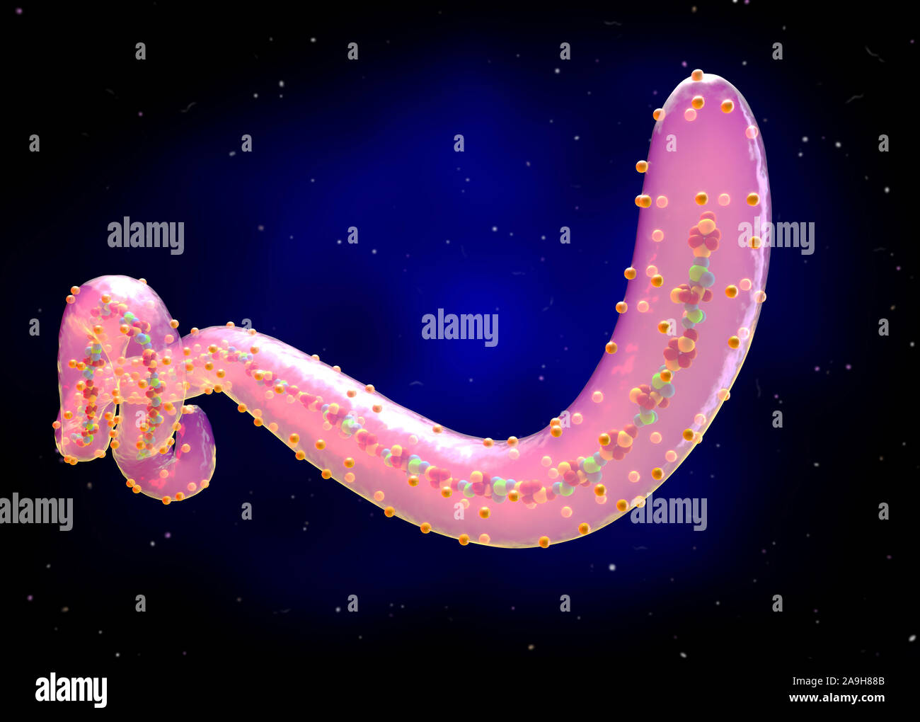
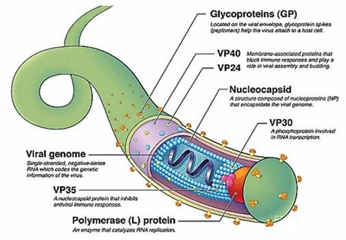
Gp2 forms the protein machinery which drives fusion of the viral membrane with the host cell. Ebola incurs mutations whenever it is copied, just as humans, plants, and other organisms introduce new mutations to their genomes each time they reproduce. The public will likely be apprehensive, frightened or anxious, and require information and reassurance. The number of ebola cases occurring in maine will be relatively small. Results crystal structure of ebov vp35 was solved to high resolution. Ebov has a striking, filamentous structure: 1 b and c).in the crystal, there are 2 nearly identical monomers (a and b) of vp35 iid in the asymmetric unit [supporting information (si) text. Ebola virus biology and research. An ebola case(s) occurring within the state of maine will be considered a public health emergency. Four of the six known ebolaviruses, including ebov, cause a severe and often fatal hemorrhagic fever in humans and other mammals, known as ebola virus disease (evd). The three gp1 subunits (colored blue and green), mediate attachment to new host cells, and are tethered together by the three gp2 subunits (white). The figure above is an organizational chart showing the ministry of health and social welfare (mohsw) ebola response framework in liberia during july 2014. The helical nucleocapsid acquires an envelope by budding from the plasma membrane, a process driven by the vp40 matrix protein.
The three gp1 subunits (colored blue and green), mediate attachment to new host cells, and are tethered together by the three gp2 subunits (white). Ebola virus is a highly pathogenic filovirus causing severe hemorrhagic fever with high mortality rates. Filamentous, enveloped viruses that cause haemorrhagic fever. Ebov), is one of six known species within the genus ebolavirus. Ebola virus (ebov) vp30 is a multifunctional protein that plays a role in transcription, but molecular details remain unknown.
Ebola Virus Antigens Creative Diagnostics from www.creative-diagnostics.com In humans, ebolaviruses cause fatality in 25 to 90 percent of cases. Ebola virus is a highly pathogenic filovirus causing severe hemorrhagic fever with high mortality rates. The first symptoms are usually fever, sore throat, muscle pain, and headaches. Terrible suffering in western africa has refocused the world's attention on ebola viruses, for which there is no vaccine or cure. They are cylindrical/tubular, and contain viral envelope, matrix, and nucleocapsid components. Ian michelow, who has studied an approach. Filovirus particles form long, sometimes branched, filaments of varying shapes, as well as shorter filaments, and may measure up to 14,000 nanometers in length with a diameter of 80 nanometers. Ebov has a striking, filamentous structure:
Epidemic of ebola hemorrhagic fever which appeared in the countries of west africa in 2014, is the largest outbreak which occurred so far.
The ebola virus is the causative agent for ebola hemorrhagic disease in humans and in other mammals. It is caused by an infection with one of five known ebola virus species, four of which can cause disease in people. If scientists can decipher a protein's structure, they can then identify drugs able to disrupt its activity. Ministry of health and social welfare ebola response framework — liberia, july 2014. Results crystal structure of ebov vp35 was solved to high resolution. Evolution occurs when these mutations are passed down from generation to generation and spread through a population, often via natural selection. The viruses are masters of their attack, but researchers are working hard to fight them, said dr. The number of ebola cases occurring in maine will be relatively small. Ebola virus (ebov) vp30 is a multifunctional protein that plays a role in transcription, but molecular details remain unknown. Four of the six known ebolaviruses, including ebov, cause a severe and often fatal hemorrhagic fever in humans and other mammals, known as ebola virus disease (evd). Case fatality rates have varied from 25% to 90. Epidemic of ebola hemorrhagic fever which appeared in the countries of west africa in 2014, is the largest outbreak which occurred so far. Ebola virus disease (evd), formerly known as ebola haemorrhagic fever, is a rare but severe, often fatal illness in humans.
Select by hand blue strand that is near 3csy antibody loops. The three gp1 subunits (colored blue and green), mediate attachment to new host cells, and are tethered together by the three gp2 subunits (white). Meaning it can take on many shapes. The figure above is an organizational chart showing the ministry of health and social welfare (mohsw) ebola response framework in liberia during july 2014. The crystal structure of the zaire ebolavirusgp in its trimeric, prefusion conformation (3 gp1plus 3 gp2) in complex with a neutralizing antibody fragment, derived from a human survivor of the 1995 kikwit outbreak, was recently determined.
Ebolavirus Glycoprotein Structure And Mechanism Of Entry Future Virology from www.futuremedicine.com Ebov), is one of six known species within the genus ebolavirus. Ebola incurs mutations whenever it is copied, just as humans, plants, and other organisms introduce new mutations to their genomes each time they reproduce. Ebola, also known as ebola virus disease (evd) and ebola hemorrhagic fever (ehf), is a viral hemorrhagic fever in humans and other primates, caused by ebolaviruses. Nucleoproteins from other viruses from the larger family that ebola belongs to, were also familiar to researchers. Filoviruses are within the order mononegavirales, which also includes rabies virus, measles virus, and respiratory syncytial virus. The number of ebola cases occurring in maine will be relatively small. Select by hand blue strand that is near 3csy antibody loops. Atomic structures are shown on the right, with portions that have not been determined shown with schematic circles.
Ebola virus disease (evd), formerly known as ebola haemorrhagic fever, is a rare but severe, often fatal illness in humans.
In order to be prepared to respond to all acute public health emergencies, including a potential ebola virus disease event, countries need to review and enhance national public health emergency preparedness and response plans, and national command and coordination structures. Four of the six known ebolaviruses, including ebov, cause a severe and often fatal hemorrhagic fever in humans and other mammals, known as ebola virus disease (evd). These are usually followed by vomiting. Ebola, infectious disease caused by a virus of the family filoviridae that is responsible for a severe and often fatal viral hemorrhagic fever. Terrible suffering in western africa has refocused the world's attention on ebola viruses, for which there is no vaccine or cure. Since 1976 there have been approximately 35 outbreaks of the ebola virus with the most recent outbreak infecting more than 24,000 people and causing nearly 10,000 deaths. Ministry of health and social welfare ebola response framework — liberia, july 2014. Mohsw leadership recognized the organizational structure and overall response could be further optimized and sought to. The first symptoms are usually fever, sore throat, muscle pain, and headaches. An ebola case(s) occurring within the state of maine will be considered a public health emergency. Gp2 forms the protein machinery which drives fusion of the viral membrane with the host cell. The helical nucleocapsid acquires an envelope by budding from the plasma membrane, a process driven by the vp40 matrix protein. Filoviruses are within the order mononegavirales, which also includes rabies virus, measles virus, and respiratory syncytial virus.
Symptoms typically start anywhere between two days and three weeks after becoming infected with the virus ebola. The human antibody kz52 (yellow) binds an epitope at the base of the gp chalice where it bridges gp1 to gp2.


0 Komentar